
What Is SAXS?
Small Angle X-ray Scattering (SAXS) analyses repeating nanoscale structures by measuring the scattering of X-rays at very small angles as they pass through a material. SAXS provides information about structural features such as particle size and shape, typically in the range of approximately 1 to 100 nanometers.
Statistically representative measurements
Probes millions of particles or structural features simultaneously, providing robust nanoscale characterization.
Nanoscale structural insight
Reveals particle size, shape, spacing, aggregation, and pore structure in the ~1 - 100 nm size range.
Broad material capability
Suitable for crystalline and amorphous materials, including polymers, colloids, nanoparticles, porous materials, and biological systems.
Why Use SAXS?
SAXS is used when structural information is needed, but direct imaging techniques such as electron microscopy are impractical or examine too small of a volume to be representative of the material as a whole. The technique provides statistically meaningful measurements of large sample volumes and enables characterization of internal structures in bulk materials with minimal sample preparation.
Common applications include determining nanoparticle size distributions, studying polymer morphology, analyzing pore structures in porous materials, evaluating nanocomposites, and investigating self‑assembled structures in biological or soft matter systems.
Nanoparticle sizing and shape analysis
Quantifies particle size distributions, morphology, and aggregation states in powders and liquid suspensions.
Polymer and soft-matter morphology
Measures domain spacing, phase separation, micelles, gels, and hierarchical structures in polymers and self-assembled materials.
Porous and nanostructured materials
Characterizes pore size, nanoscale ordering, and structural evolution in catalysts, membranes, battery materials and other porous systems.
How SAXS Works
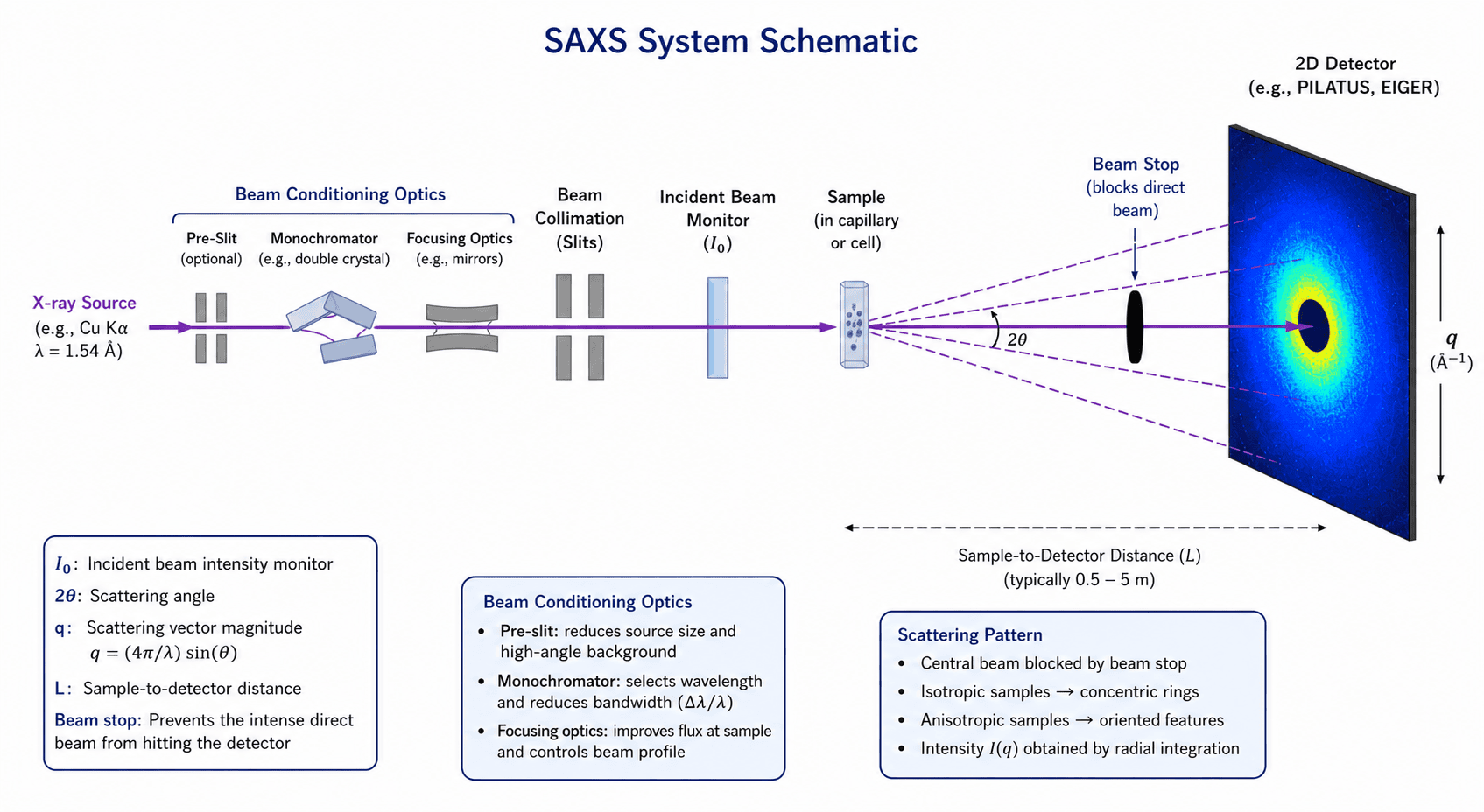
In SAXS, a monochromatic X‑ray beam is directed through a sample. Variations in electron density within the material cause the X-rays to scatter at small angles relative to the incident beam. The scattered radiation is collected by a detector, producing a scattering profile of intensity versus momentum transfer (q). Analysis of the scattering profile enables characterization of nanoscale structural features, including particle size, shape, spacing, aggregation and structural ordering.
Key Components of a SAXS System
- X-ray Source – Generates X-rays, typically Cu Kα radiation (λ = 1.5406 Å), for laboratory SAXS instruments.
- Focusing and Beam Conditioning Optics – Mirrors, multilayers, or collimation optics are used to shape, focus and define the X-ray beam.
- Aperture and Slits – Define beam size and divergence while minimizing background scattering.
- Sample – Positioned in the X-ray beam, typically in air, vacuum or controlled environment.
- 2D Detector – Records the scattered X-ray intensity over a range of scattering angles.
- Beam Stop – Blocks the intense directly transmitted beam to protect the detector and improve measurement quality.
- Sample-to-Detector Distance – A key measurement parameter that influences the accessible q-range and size scale of interest.
Laboratory SAXS instruments commonly use Cu Kα radiation, while advanced studies may utilize synchrotron radiation to achieve higher X-ray flux, improved signal-to-noise ratio, and enhanced measurement resolution.
The SAXS Measurement Process
- X-rays are generated by the source and formed into a collimated beam.
- Beam-conditioning optics shape, monochromate and define the beam.
- The beam passes through the sample, where nanoscale variations in electron density cause the X-rays to scatter.
- The scattered X-rays are recorded by a two dimensional detector as a function of scattering angle.
- The scattering data is converted into a one-dimensional scattering profile, I(q) vs. q, for analysis and structural modeling.
Scattering Vector (q)
q = (4π/λ) sin(θ)
where 2θ is the scattering angle and λ is the X-ray wavelength.
Typical SAXS measurements cover a q range of approximately 0.01–5 nm⁻¹, corresponding to real-space structural features ranging from roughly 1–100 nm.
Equipment Used for SAXS
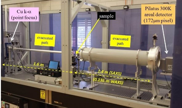
Covalent Connect leverages a network of academic and commercial partner laboratories to access SAXS instrumentation best suited to each application. Shown below is the SAXS system at the University of Illinois at Urbana-Champaign, one of our lab partners.
Equipment Specs
Radiation / wavelength: Cu K-alpha, 0.15418 nm
Main optics: Genix3D ULD microfocus source including focusing mirror (beam size at sample: 0.8 x 0.8 mm2), evacuated primary and secondary beam paths, sample stage with theta (angle of incidence) rotation and horizontal/vertical sample translation; high-speed Pilatus 300 detector (172 microns pixel size, 500 eV resolution) for 2D areal data acquisition.
Key Differentiators
Primary outputs from SAXS measurements include scattering intensity as a function of scattering vector (I(q)), particle size distributions, radius of gyration (Rg), correlation lengths, and information related to particle shape, aggregation, and structural ordering.
SAXS is sensitive to nanoscale features ranging from 1–100 nm. Typical sample formats include millimeter‑scale solids, thin films, powders and liquid samples contained in capillaries. The technique is compatible with a wide range of materials including polymers, nanoparticles, colloidal dispersions, porous materials, biological systems and bulk materials.
Successful measurements require sufficient electron density contrast between structural features of interest and surrounding material. Samples should be representative of the material being studied and free from large aggregates or defects that can distort the scattering signal.
Strengths
• Sensitive to nanoscale structures (1–100 nm)
• Non‑destructive measurement technique
• Minimal sample preparation required
• Applicable to solids, liquids, gels, and thin films
• Provides statistically representative structural information from large sample volumes, compared to microscopy techniques like TEM
• Effective for studying particle size distributions, aggregation behavior, and nanoscale ordering
Limitations
• Requires significant electron density contrast between phases
• Provides indirect structural information that typically requires data modeling and interpretation.
• Lower spatial resolution compared with imaging techniques such as TEM
• Data interpretation can be complex for multi‑component systems
• Accurate background subtraction and instrument calibration are required
Example Outputs
Typical SAXS results include plots of scattering intensity versus scattering vector (I(q) vs q). Analysis of these curves can provide information on particle size distributions, radius of gyration (Rg), fractal behavior, structural ordering, and characteristic length scales within a material.
Representative theoretical SAXS plots are shown below to illustrate how scattering features are interpreted and related to material structure.
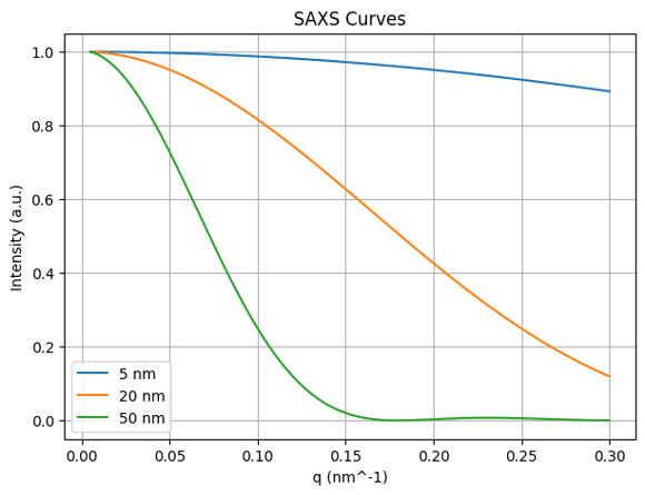
Plots intensity versus scattering vector (q). Larger particles produce features at lower q values because size and reciprocal-space position are inversely related.
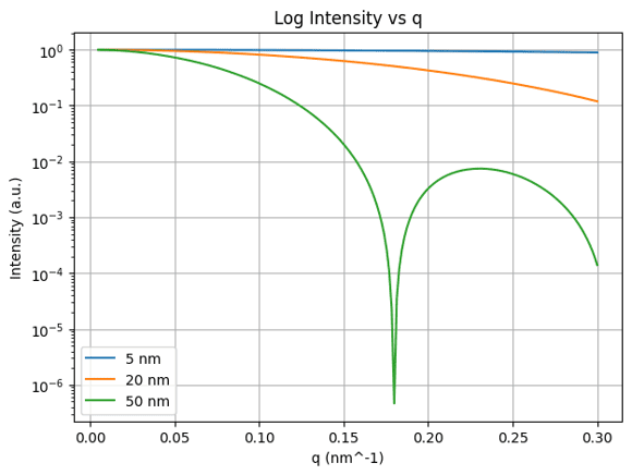
Semi-Log SAXS Plot
A common publication format. The logarithmic intensity axis makes weak scattering features easier to observe.
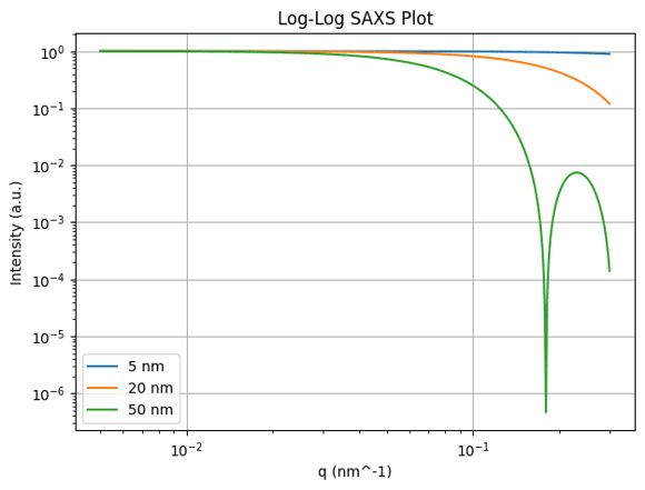
Log-Log SAXS Plot
Used to identify Guinier, power-law, fractal, and Porod scattering regimes. Slope analysis often provides clues regarding morphology.
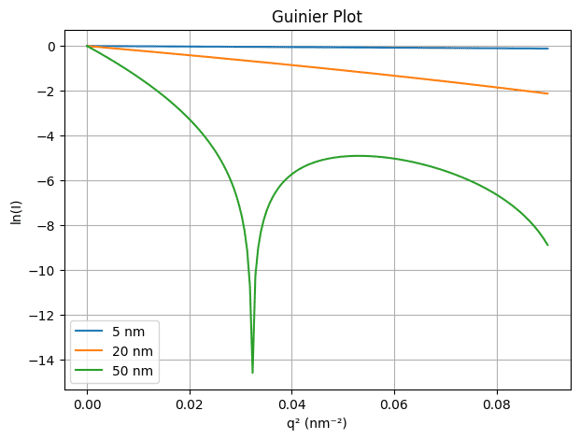
Guinier Plot
The low-q region becomes approximately linear when plotting ln(I) versus q². The slope can be used to estimate the radius of gyration (Rg).
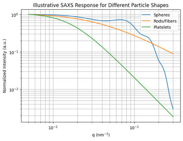
Particle Shape Comparison
The image above shows illustrative scattering behavior for spheres, rods/fibers, and platelets. Different shapes produce characteristic scattering slopes and form-factor features.
How Shape Affects a SAXS Pattern
Spheres
Spherical particles exhibit characteristic form-factor oscillations, with oscillation positions related to particle size. Because their scattering behavior is well understood, spherical particles are often the most straightforward morphology to model quantitatively.
Rods / Fibers
Rod-like structures typically exhibit a slower intensity decay at intermediate q and may show an approximate q⁻¹ dependence over a characteristic scattering range. Examples include nanotubes, nanowires, fibrils, and elongated precipitates.
Platelets / Sheets
Plate-like structures often exhibit enhanced low-q scattering and may show an approximate q⁻² dependence over an appropriate q range. Examples include graphene, clays, layered materials, and thin nanosheets.
Rule of Thumb
Morphology | Approximate Slope |
Rods/Fibers | q⁻¹ |
Platelets/Sheets | q⁻² |
Compact particles | q⁻⁴ (Porod) |
Fractal aggregates | Between q⁻¹ and q⁻⁴ |
Shape interpretation from slope alone can be misleading because aggregation, polydispersity, surface roughness, and hierarchical structures can generate similar scattering behavior.
Recommended Workflow
1. Determine the Guinier region and estimate Rg.
2. Evaluate intermediate q behavior for morphology and structural information.
3. Analyze the Porod region for interface and surface characteristics.
4. Fit appropriate structural models to extract quantitative parameters.
5. Validate results with complementary techniques such as TEM or SEM when available.
SAXS Applications
Nanoparticle characterization
Determination of particle size, size distributions, and aggregation behavior.
Polymer morphology
Analysis of phase separation, domain spacing, and hierarchical structure.
Porous materials
Characterization of pore size, pore structure, and nanoscale ordering.
Nanocomposites
Evaluation of filler dispersion, particle interactions, and structural organization.
Self-assembled materials
Investigation of block copolymers, micelles, and other self-assembled structures.
Biological macromolecules
Structural characterization of proteins, protein complexes, and other biological assemblies in solution.
Thin films and multilayers
Analysis of nanoscale structure, layering, and periodicity.
Techniques That Complement SAXS
Provides direct imaging of particle size, shape, defects, interfaces and local morphology.
Provides rapid particle size measurements in liquid suspensions and can identify aggregation or agglomeration.
Determines crystal structure, phase composition, crystallinity, and crystallite size.
Measures thin-film thickness, density, interface quality and surface roughness.
Produces real-space measurements of surface topography, roughness and nanoscale surface features.
Provides high-resolution imaging of particle morphology, surface features, and microstructure over larger fields of view than TEM.
Frequently Asked Questions
What sample types can be analyzed by SAXS?
SAXS can analyze a wide range of materials including nanoparticles, polymers, catalysts, battery materials, semiconductor materials, thin films, porous materials, biological macromolecules, and colloidal suspensions. Samples can be analyzed as powders, liquids, gels, thin films, or bulk solids.
What is the typical size range measurable by SAXS?
SAXS is most sensitive to structural features between approximately 1 and 100 nm, although the accessible size range depends on instrument configuration and measurement conditions. Structures smaller than 1 nm are typically better characterized by WAXS or XRD, while structures larger than 100 nm may require USAXS, microscopy, or light scattering techniques.
How does SAXS compare to TEM?
SAXS and TEM are complementary techniques. SAXS measures the average structure of a large sample volume and provides statistically robust size distributions. TEM directly images individual particles and reveals morphology, defects, and interfaces. SAXS is generally better suited for obtaining statistically representative particle size distributions, while TEM excels at visualizing particle morphology and validating structural interpretations.
Is SAXS destructive?
No. SAXS is a non-destructive characterization technique that typically requires minimal sample preparation. In many cases, samples can be recovered and analyzed further using complementary methods.
What standardized test methods use SAXS?
The following test methods use SAXS:
- ISO 17867 – Particle Size Analysis by Small-Angle X-ray Scattering (SAXS). Primary international standard for nanoparticle sizing using SAXS.
- NIST SAXS Reference Materials and Calibration Guides – Reference materials and best-practice guidance for instrument calibration, validation, and uncertainty analysis.
- IUCr Biological SAXS Guidelines – Reporting and data-quality recommendations for solution SAXS and structural biology studies.
- SEMI GISAXS/CD-SAXS Practices – Semiconductor-industry guidance for nanostructure and critical-dimension metrology using scattering techniques.
- ISO 22412 – Dynamic Light Scattering (DLS). Frequently used alongside SAXS for nanoparticle sizing and comparison studies.
- ISO 13318 – Nanoparticle Tracking Analysis (NTA). Often referenced when comparing nanoparticle sizing methods with SAXS.